Specimen imaging
High-magnification, focus-stacked documentation of botanical, arthropod and horticultural specimens with controlled lighting and consistent capture.
Request a quote →Scientific imaging · British Columbia
Publication-quality focus-stacked imagery of plant, arthropod and small biological specimens, built for scientific detail and clear visual communication.

Services
From a single specimen to a publication figure set, each project begins by defining the structure, scale and intended use.
High-magnification, focus-stacked documentation of botanical, arthropod and horticultural specimens with controlled lighting and consistent capture.
Request a quote →Capture, stack cleanup, calibrated scale bars, panel layout and caption-ready technical metadata for manuscripts, posters and reports.
Plan a figure →Workflow design, equipment selection, rail-step planning, lighting refinement and diagnosis of haze, movement and magnification-breathing artifacts.
Book a consultation →Rights-managed scientific and editorial images for books, articles, exhibits, teaching materials, covers and science communication.
License an image →Selected work
Each final image combines a sequence of narrow focal planes while preserving the specimen’s three-dimensional form.
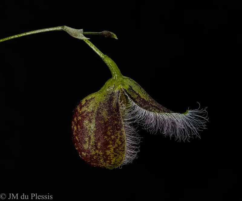
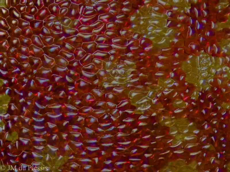
Case study · Orchid morphology
A coordinated image series can establish overall form, reveal diagnostic structures and document fine surface texture—without flattening the subject into a conventional slide preparation.
Client proof · Multi-scale series
Two delivered series presented proof-sheet style — every frame focus-stacked, caption-ready and referenced by its catalogue ID, from whole-flower habit to cellular surface.
Series can be licensed frame-by-frame or as a coordinated set — every image carries its capture record and rights terms.
License a frame from these series →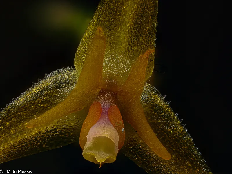 Specimen study · 4 composites
Specimen study · Lepanthes myiophora
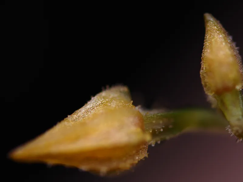
A complete record of one plant: face-on at 2.5× for display, from below at the same scale, from slightly above at 5×, and as a bud before anthesis — four focus-stacked composites, licensable frame by frame or as a coordinated set.
View the myiophora series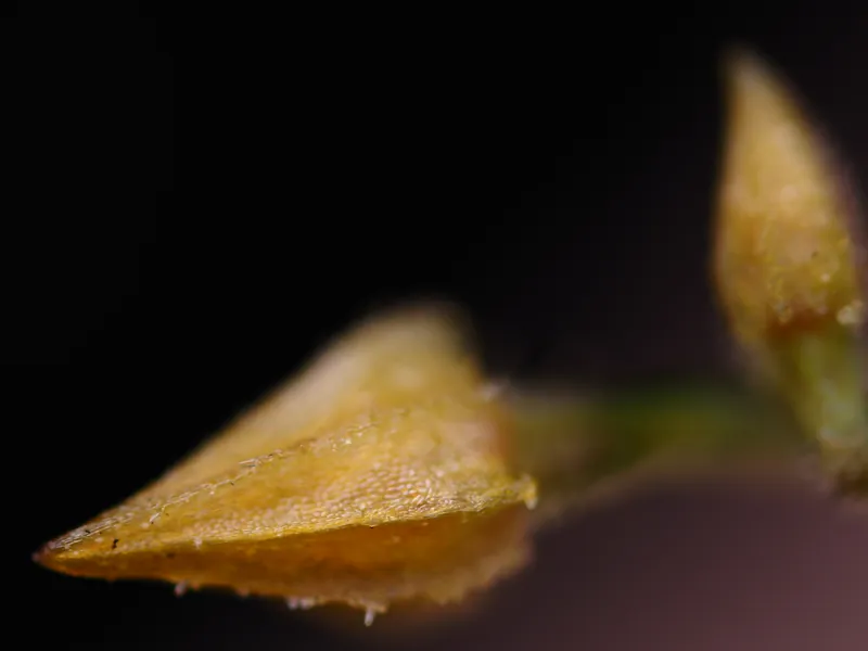
Method
At high magnification, depth of field collapses to a thin optical section. An automated rail advances the camera through the specimen, recording overlapping planes that are merged into an extended-depth composite.
Technical notes
Practical articles developed from repeated high-magnification capture, failure analysis and workflow refinement.
The plain-language entry point to the series: what macro actually means, why depth of field collapses at high magnification, and how focus stacking rebuilds it.
Read note ↗Why defocused foreground structures contaminate sharp regions—and why stacking software cannot reconstruct detail that was never recorded cleanly.
Read note ↗How small physical shifts become large sensor-plane displacements and produce doubling, ghosting and unresolved transitions.
Read note ↗Understanding apparent scale changes across a stack and the demands they place on alignment and composite integrity.
Read note ↗Why vibration is the least visible and least repairable artifact in deep stacks, and the control hierarchy that prevents it.
Read note ↗Choosing step size from 2.5× to 10×: depth-of-field collapse, the 80 percent rule, and worked values for real configurations.
Read note ↗Two optical worlds with one goal — extended depth of field — compared honestly: specimen handling, aperture arithmetic, resolution ceilings, and when each tool wins.
Read note ↗Image catalogue
Reference the image ID when requesting editorial, educational, exhibit, cover or commercial use.
Rights-managed licensing
Licences can be tailored by medium, territory, duration, placement and exclusivity. Full-resolution masters and caption-ready metadata are supplied after agreement — originals are typically 10368 × 7776 or 5184 × 3888 pixels, and PNG or TIFF masters can reach hundreds of megabytes, so the website displays optimized previews only.
Browse the licensing catalogue
About
Jean-Mornay du Plessis is a plant scientist and scientific photographer working across morphology, controlled-environment horticulture and high-magnification imaging.
Based in British Columbia, his practice combines specimen knowledge with precision optical workflows to produce images for research, teaching, publishing and public communication.
Start a project
Share the specimen, approximate size, scientific question, intended use and deadline. A scoped response will follow.